



Estimated reading time: 4 minutes
A life-threatening blood clot in the lungs can leave clinicians facing difficult treatment decisions. Medications used to dissolve clots can take hours to work and carry bleeding risks, while procedures to remove them can damage blood vessels.
Researchers at the University of Michigan are developing a new approach that could eventually offer another option for treating pulmonary embolism, the third leading cause of cardiovascular death in the United States.
Backed by a $3.74 million grant from the National Institutes of Health (NIH), the five-year project will investigate whether a catheter-based device that delivers swirling ultrasound waves can break apart dangerous blood clots more quickly and safely than existing treatments.
In early laboratory testing, researchers restored blood flow through a three-inch-long clot in just eight minutes using cow’s blood and artificial blood vessels. The technology has not yet been tested in humans.
Want to see more Nurse Approved articles in your Google results? Add us as a preferred source.
Add Nurse Approved as apreferred source on Google
Why Pulmonary Embolism Treatment Remains Challenging
Pulmonary embolism occurs when a blood clot, often originating in the legs, travels to the lungs and blocks blood flow. Large clots can interfere with oxygen exchange and place sudden strain on the heart, making severe pulmonary embolism a medical emergency.
According to the University of Michigan researchers, clot-dissolving medications can take up to 24 hours to work, may fail in approximately one-third of cases, and carry a risk of serious internal bleeding.
Catheter-based mechanical clot removal offers another approach, but the procedure can damage fragile blood vessel walls. The most severe cases may require open-chest surgery.
These limitations have prompted researchers to explore ways to restore blood flow faster while reducing treatment-related complications.
How Swirling Ultrasound Could Break Up Blood Clots
The experimental device uses a technique called vortex ultrasound, which produces twisting sound waves designed to disrupt a blood clot’s internal structure.
A miniature ultrasound source mounted at the tip of a catheter would deliver the waves directly to the clot. Researchers believe the swirling waves create bubbles within the clot, helping break it apart without damaging surrounding blood vessel walls.
Delivering ultrasound from inside the vessel could also overcome a limitation of external ultrasound treatment. Lung structures can interfere with sound waves, making it difficult to target clots accurately.
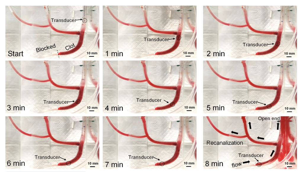
Preliminary research suggests vortex ultrasound could potentially break up clots in as little as 30 minutes.
In the team’s earlier proof-of-concept experiment, an ultrasound transducer moved through a three-inch clot inside plastic tubes designed to mimic blood vessels. The procedure restored blood flow without damaging the artificial vessel walls.
Although the laboratory results are promising, researchers still need to determine whether the device can safely and effectively treat pulmonary embolisms in patients.
“With this approach, we hope to treat clots in the lungs more quickly and safely while potentially providing an alternative to open-chest surgery for severe pulmonary embolism,” said Chengzhi Shi, PhD, associate professor of mechanical engineering at the University of Michigan and principal investigator of the project.
What the Research Could Mean for Nurses
For emergency and critical care nurses, pulmonary embolism is a time-sensitive condition in which changes in oxygenation and cardiovascular status can signal serious deterioration.
Nurses caring for patients with severe pulmonary embolism must monitor for clinical changes while patients undergo evaluation and treatment, including complications associated with clot-dissolving medications or catheter-based procedures.
If future studies establish that vortex ultrasound is safe and effective in humans, it could expand treatment options for patients with severe pulmonary embolism.
However, researchers have not established whether the technology reduces bleeding complications, prevents blood vessel injury, or improves patient outcomes compared with existing treatments.
The early findings are promising, but they do not yet support changes in clinical practice or nursing care.
Five Years of Research Ahead
The NIH grant will support five years of development and testing to prepare the technology for potential human clinical use.
Researchers plan to refine the catheter design, determine the most effective shape and frequency of the ultrasound waves, and evaluate short- and long-term safety in pigs.
The project brings together engineers and vascular surgeons from the University of Michigan and North Carolina State University.
For now, vortex ultrasound remains an experimental approach to one of cardiovascular medicine’s most urgent challenges: restoring blood flow quickly while minimizing treatment-related risks for patients facing a potentially fatal emergency.
